A Baby Girl Was Born With a 5.7-Centimeter ‘Tail’—But Doctors Had to Look Beneath the Skin Before Removing It
The baby girl was born at full term after an uncomplicated pregnancy. During her examination, doctors found something they rarely encounter: a soft, skin-covered appendage measuring 5.7 centimeters, extending from her lower back near the tailbone.
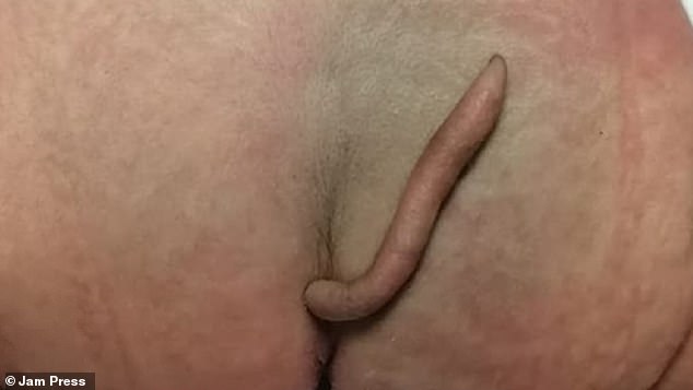
It looked like a small tail. But its appearance could not tell the medical team everything they needed to know.
Before deciding how to remove it, they had to find out whether the structure was connected with a deeper abnormality of the spine or spinal cord. That question would determine the meaning of the finding and the approach to her care.
The case, reported by Josue Rueda and colleagues in the January 2022 issue of the Journal of Pediatric Surgery Case Reports, involved a newborn in northeastern Mexico. Her identity was not published. The paper recorded that her parents gave informed consent for the case to be shared.
When news reports brought the story to a wider audience later in 2022, the unusual photograph naturally attracted attention. The medical account, however, contains a more reassuring story than that first image might suggest.
An unusual finding after an ordinary pregnancy
According to the case report, the girl was delivered by Caesarean section and initially appeared healthy apart from the appendage. Her prenatal history did not identify complications that had pointed doctors toward this finding before birth.
The structure emerged slightly to the left of the midline in the sacrococcygeal region, near the bottom of the spine. It was slender, covered by skin and fine hair, and narrowed toward its tip.
The team documented that it could be moved gently by an examiner without causing pain. It did not move by itself. They also recorded a response to a sensation test, showing that it contained sensitive tissue.
These observations helped describe what was present, but they were only the beginning of the assessment. A visible appendage in this region can have different underlying explanations. Its outline alone is not enough to establish which one applies.
Why the scans mattered more than the photograph
The paper discusses several ways clinicians classify so-called human tails. Some definitions distinguish a soft-tissue appendage from a tail-like projection associated with another structural condition. The same appearance can therefore lead to very different medical questions.
For this baby, the team investigated the structures underneath. An X-ray did not show bone within the appendage. Further assessment included magnetic resonance imaging, which the authors reported did not identify the spinal abnormalities they were looking for.
The report also described reassuring findings from other examinations, including screening related to her heart and hearing and assessment of her urinary system.
That is the important turn in the story. The striking external finding had brought the child to medical attention, but the work beneath the surface helped establish a favorable outlook.
The authors emphasized imaging because a tail-like appendage can sometimes be a marker of an underlying spinal condition. Their conclusion was not that every similar-looking structure is harmless or can be treated in the same way. It was that assessment had shown why a relatively straightforward operation was appropriate in this particular case.
The operation came after reassessment
The team did not remove the appendage immediately after birth. The baby was followed up, and at two months old she was reassessed by paediatric and surgical clinicians.
Her weight gain and growth were appropriate for her age. The appendage had also grown, increasing by 0.8 centimeters to approximately 6.5 centimeters. Those measurements describe two different points in her early life: 5.7 centimeters at birth and 6.5 centimeters at removal.
Once the assessment had ruled out the underlying spinal abnormalities described in the report, the surgeons removed the tissue and closed the area using a local reconstructive technique called Limberg plasty.
The paper described an outpatient procedure under local anaesthesia with sedation. It did not report an emergency operation or a life-saving intervention. The distinction matters because the baby’s unusual appearance was not, by itself, evidence that she was critically ill.
The removed tissue was examined. The findings included soft tissue, blood vessels and nerve bundles covered by skin, without the pathological abnormalities the authors described looking for.
The child was discharged, and the published account reported no complications at follow-up. That is the outcome available in the medical record; it does not supply a later public account of her school years or current life.
A rare case with a reassuring ending
The authors believed this was the first published description of the finding in a Mexican newborn, to the best of their knowledge. That is a statement about the medical literature available to them, rather than proof that no similar child had ever been born in the country.
It is also important to distinguish the publication date from the date of birth. The report appeared in January 2022 and received wider news coverage in November that year. Those dates do not establish the baby’s exact birthday.
What the report does establish is a sequence of careful decisions: an unusual finding at birth, imaging to assess deeper structures, reassessment as the child grew, and removal after the team had the information needed to proceed.
The photograph explains why people noticed the story. The medical work explains why it ended as reassuringly as it did.
For the anonymous little girl at its center, the final lines were the most welcome ones: she had been discharged, and no complications were reported at follow-up.
A rare physical feature had made her case worthy of publication. Her clinicians’ aim was to understand it, treat it appropriately, and allow her care to return to the ordinary business of growing.
Clinical photograph reproduced in contemporary reporting; visible image credit: Jam Press. Case report: Rueda and colleagues, Journal of Pediatric Surgery Case Reports, 2022.